|
2.2.2 THE INTERNAL SEX ORGANS
The female internal sex organs consist of the ovaries, the Fallopian tubes, the uterus, and the vagina.
The Ovaries
The ovaries (the female sex glands or gonads) are two walnut-sized bodies which are located inside the abdomen on either side of the uterus. The ovaries serve a double function:
They produce eggs which are released into the Fallopian tubes.
They produce hormones which are secreted directly into the bloodstream.
The Production of Eggs
Before a baby girl is born, all of the cells that will later grow into eggs are already formed in her ovaries. In their primitive beginnings, the cells are called oogonia. These oogonia turn into primary oocytes, some of which eventually give rise to mature eggs (ova). The process of egg production; called oogenesis, begins in the female fetus, but soon comes to a halt at birth. Thus, every girl is born with nearly 500.000 primary oocytes which remain in their state of suspended development until she reaches puberty. (During this time no new oocytes are produced. On the contrary, most of those that had developed earlier gradually die. By the time a girl reaches puberty, there may be no more than 30,000 primary oocytes left that are capable of further development. At the age of thirty, this number has dwindled even further to about 10,000, and when the woman reaches her menopause, all primary oocytes are gone.) Once the process of oogenesis has resumed during puberty, one or several mature eggs are produced each month by either one of her ovaries until both of them cease functioning following menopause. In the course of her fertile life, a woman may produce some 400 mature eggs. Of course, only a very small fraction of these can ever contribute to conceptions.
All of this provides a striking contrast to the way sperm cells are produced in the male (continuous production of millions of sperm daily, beginning with puberty; for details, see "The Male Sex Organs.").
The development of a mature egg proceeds in several steps: Each primary oocyte is contained in a cluster of supporting cells. These clusters lie beneath the outer layer of the ovary. Each month, under the influence of certain hormones, one of the clusters grows to a point where it appears as a rather large blister on the surface of the ovary. This blister is called a Graafian follicle {after the 17th-century anatomist de Graaf). During the period of follicle growth, the primary oocyte, which like any other female body cell contains 46 chromosomes (including two X chromosomes), divides into two new cells of very unequal size: a relatively large secondary oocyte and a minute so-called polar body. In this division, the 46 chromosomes are split apart, and half of them are allotted to each of the new cells. Thus, the secondary oocyte as well as the polar body each contain only 23 chromosomes (including one X chromosome).
The polar body dies and disintegrates. Only the secondary oocyte is destined for further maturation. First, it floats freely inside the growing follicle which contains fluid. Eventually, the follicle bursts, releasing the secondary oocyte into the abdominal cavity. This release is known as ovulation. The secondary oocyte then enters the nearest Fallopian tube. Here it divides again into two new cells of unequal size: a relatively large ootid (mature ovum) and a minute second polar body. However, this time the division reproduces rather than splits the number of chromosomes. Thus, both of the new cells retain 23 chromosomes (including one X chromosome). This last division and the expulsion of the second polar body occur only after fertilization. While the second polar body dies just as the first one, the 23 chromosomes of the ootid unite with the 23 chromosomes of the spermatozoon, thus forming a new cell (the zygote) which again contains 46 chromosomes like all other cells of the body. (For details, see "Conception.")
The Production of Hormones
As described in a previous section, the female and male gonads (ovaries and testicles) also produce certain hormones. These gonadal hormones have been divided into female hormones (estrogens) and male hormones (androgens). However, these terms are somewhat misleading because both "female" and "male" hormones can be found in every female and male body. It is only the quantity of these hormones that differs. Before puberty, the estrogen and androgen levels in girls and boys are nearly equal. Then, during adolescence, the balance begins to shift. In the female body the estrogens rise to a much higher level than the androgens, and in the male body the androgens rise to a slightly higher level than the estrogens. In the female, the increase of estrogens during puberty helps to produce the secondary female sexual characteristics. (In the male, the increase of androgens helps to produce the secondary male sexual characteristics.)
In addition to estrogens (and androgens), the ovaries of a sexually mature female also produce a hormone called progesterone. The production of progesterone takes place mainly in association with the corpus luteum {Latin: yellow body) which is formed from the wall of the ruptured follicle after ovulation. During a women's fertile years, the estrogens as well as progesterone play an important role in her reproductive cycle, (For details, see "The Menstrual Cycle.")
There is still much to be learned about the nature and function of hormones in the human body. Nevertheless, a few basic facts have already been established: While the gonadal hormones are necessary for a young person's physical maturation, they are not essential for the continued sexual activity of adults. In other words, females and males need their gonadal hormones during adolescence to develop their full sexual potential. However, once the potential has been attained, they can function sexually without these hormones. Therefore, a woman who approaches menopause does not have to fear that she will lose her sexual responsiveness. Even when her ovaries have ceased their hormone production, she can continue her sexual activities as before. The same is also true, of course, for a woman who has to have her ovaries surgically removed for reasons of illness. (For details, see "The Role of Hormones.")
The Fallopian Tubes
The Fallopian tubes (named after the 16th-century anatomist Fallopius) lead from the ovaries to the uterus. They are also sometimes called oviducts (Latin: paths of eggs), a term that accurately describes their function. They provide a passageway for the egg down to the area where it could implant in case of a fertilization. (They also provide a passageway for sperm cells swimming upward from the uterus trying to reach the egg.) The wide ovarian end of a Fallopian tube has finger-like extensions called fimbriae (singular: fimbria) which move across the surface of the ovary; the uterine end leads directly into the inside of the uterus.
The fertilization of an egg normally occurs in the upper part of a Fallopian tube. Inside the tube, there are innumerable hair-like growths called cilia (singular: cilium) whose movements, together with muscular contractions of the tubal wall, sweep the egg toward the uterus. (Inside the male vas deferens, the sperm cells are transported the same way since they are still unable to move by themselves at that point.)
The Uterus
The uterus (Latin: womb) is a muscular organ which is situated between and slightly below the ovaries, approximately in the center of the lower abdomen. The shape of the uterus, which is about 3 inches long, resembles that of a small pear turned upside down. The Fallopian tubes enter the uterus on either side near the top. The wide upper part, known as the body of the uterus, is usually tilted forward over the dome of the urinary bladder, and it is separated from the narrow lower part by a slight constriction. This lower part is called the cervix or neck of the uterus, and it ends in the deep portion of the vagina. The cervix contains a small opening through which sperm cells can travel from the vagina into the uterus. However, except for a certain period during ovulation, the cervical opening is plugged by an impenetrable mucus.
The thick walls of the uterus are made up of three layers: the external cover called the perimetrium, the middle or muscular layer called the rnyometrium, and the inner layer called the endometrium. This endometrium consists of special tissue which thickens every month as the uterus prepares for the possible implantation of a fertilized egg. (Also see "Conception.") If no implantation occurs, the endometrium deteriorates and is discharged through the cervix and the vagina during menstruation. (See "The Menstrual Cycle.")
In case of a pregnancy, the uterus expands with the growing fetus. The extraordinary muscular structure of the myometrium not only allows for such vast expansion, but also provides the necessary pressure during labor when the fetus is finally expelled. (See "Birth.")
The uterine muscles also contract during orgasm. (For details, see "The Female Sexual Response.")
The Vagina
The vagina (Latin: sheath) is a muscular tube about 31/2 inches long extending from the cervix to an external opening which is part of the vulva. The vagina serves three main functions:
It provides a passageway for the menstrual flow from the uterus to the outside. (See "The Menstrual Cycle.")
It serves as a receptacle for a man's penis and his ejaculated sperm which then may move on through the cervix (see "Conception.").
It provides a passageway for the baby during birth from the uterus to the outside. (See "Birth.")
Under ordinary circumstances, the vagina is a collapsed tube, i.e., more a potential than actual space. Its inner surface, like that of the mouth, hosts different kinds of organisms which live in a healthy ecological balance. This balance can be upset, however, as a result of chemical interference. For this reason, vaginal sprays and douches should be avoided. The vagina cleanses itself with its own secretions. It also possesses a special protection against infection. (For vaginal infections and infestations, see "Venereal Diseases.")
The vaginal walls, which lie close together, contain mucous crypts and many blood vessels, but no glands and few nerve endings. (See also "The Female Sexual Response.") During sexual excitement, these walls secrete a watery substance which serves as a lubricant during coitus. Without such lubrication, the insertion of a penis could be painful to both the woman and the man. (See "Pain During Sexual Intercourse.")
Some women also expel some fluid from the urethra during orgasm. In the past, it was often assumed that the fluid could only be urine, and this caused the women some embarrassment. Sometimes another explanation was given: The fluid must have come from the vagina itself, perhaps as sudden excessive lubrication or as secretion from the Bartholin's glands. However, both explanations were wrong: The fluid did indeed come from the urethra, but it was not urine. Recent research suggests that the fluid is secreted by a system of urethral (or paraurethral) glands, i.e., glands which surround the urethra and open into it. This system of glands is more developed in some women than in others. In any case, it corresponds to the prostate in males, which also surrounds the urethra. Some researchers have therefore gone so far as to speak of a "female prostate." By the same token, the expulsion of fluid from this glandular system during orgasmic contractions has been called "female ejaculation" especially since the fluid itself has been shown to be similar to male prostatic fluid. (There are no sperm cells in these "female ejaculations", of course.)
While only relatively few women "ejaculate" in this sense, many more (perhaps all) have a certain sensitive spot, a cluster of tissue surrounding the urethra, which can be felt and stimulated through the anterior vaginal wall. This tissue (which is probably identical or at least connected with the system of urethral glands) first swells under intense stimulation and then contributes to a specially intense orgasmic release. Anatomically, this sensitive area has now become known as the "Gräfenberg spot," after Ernst Gräfenberg who first described it in 1950.
The vagina adjusts to the size of any inserted penis, large or small. However, that portion of the vagina which lies closest to the external opening may, in some cases, become too relaxed for the preference of either sexual partner. This can happen after childbirth, for instance, or simply as a result of the aging process. Conversely, it is also possible for the vaginal entry to become so tense and tight that it cannot be penetrated. Such a vaginal spasm is called vaginismus. In either case, it should be remembered that a woman can attain a great deal of control over the function of her vaginal muscles, and that they can be developed by appropriate exercises. Some of these exercises, the so-called Kegel exercises, can easily be performed at all times, anywhere. They are described in another section of this book. (See "Sexual Dysfunction in WomenAbsence of Orgasm.")
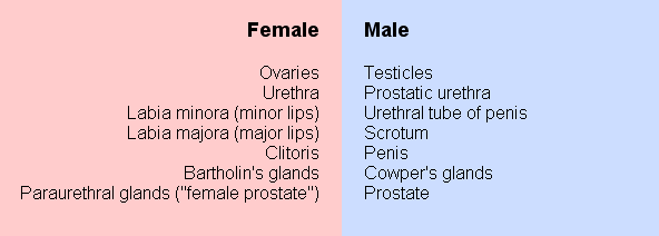
SOME HOMOLOGOUS STRUCTURES IN THE FEMALE AND MALE SEXUAL SYSTEMS
Before a baby boy is born, testosterone (a male hormone) transforms his originally undifferentiated embryo, leading to the development of a male body with male sex organs. In case of a baby girl, the absence of testosterone at this developmental stage lets the embryo grow "automatically" into a female body with female sex organs. However, since both male and female sex organs derive from the same embryonic cell mass, they still correspond to each other or, in scientific language, are "homologous."
 |
|